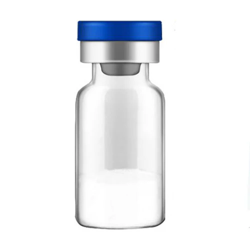
IGF-1 LR3
The Muscle Growth Accelerator
Maximize Muscle Gains: Unlock the Power of IGF-1 LR3

IGF-1 LR3, or Insulin-like Growth Factor 1 Long Arg3, is a modified version of the naturally occurring hormone IGF-1, which plays a crucial role in human growth and development. This engineered variant of IGF-1 includes an extension of 13 amino acids at the N-terminus and a substitution of Arginine (Arg) for Glutamic acid (Glu) at the third position, hence the name “Long Arg3.”
The modifications enhance the molecule’s biological activity in several ways. Firstly, IGF-1 LR3 has a reduced affinity for IGF-binding proteins, which normally regulate the biological availability and activity of IGF-1 by binding to it. This reduced binding to the proteins allows IGF-1 LR3 to remain active in the body for a longer period compared to natural IGF-1.
Secondly, by staying active longer and being less bound by these proteins, IGF-1 LR3 can more effectively stimulate the signaling pathways that lead to muscle growth and repair, cell regeneration, and other growth-related effects. This makes IGF-1 LR3 particularly interesting for research in fields related to aging, muscle development, and metabolic diseases.
Potential Benefits Under Research
- Promotion of Cell Division and Tissue Regeneration: IGF-1 LR3 is believed to enhance cell proliferation and tissue regeneration, which is beneficial for healing wounds and repairing tissues after injury.
- Muscle Growth and Myostatin Inhibition: The modified hormone may significantly impact muscle development by reducing the activity of myostatin, a protein that limits muscle growth. This could help in treating muscle degeneration and enhancing muscle repair.
- Diabetes Management: IGF-1 LR3 has potential benefits in diabetes care by improving insulin sensitivity and glucose metabolism, which could help in better management of blood sugar levels.
- Longevity and Anti-Aging: There is ongoing research into the role of IGF-1 LR3 in extending lifespan and delaying the aging process. By promoting cellular health and reducing cellular senescence, it may help maintain youthfulness and slow aging.
- Glucocorticoid Signaling Modulation: The compound could modulate glucocorticoid signaling, which is crucial in managing stress and metabolic balance. This modulation may help protect against stress-related diseases and improve metabolic health.
Dosing Protocol for Research Purposes
- 50mcg injected subcutaneously in the morning or 10-30 minutes before a workout, and within a “10 days on, 4 weeks” off cycle.
Overview
IGF1-LR3 is a modified version of insulin-like growth factor-1. The full name of the peptide is insulin-like growth factor-1 long arginine 3. All lGF-1 derivatives play prominent roles in cell division, cell proliferation, and cell-to-cell communication. Though it has similar effects, IGF-1 LR3 does not adhere to IGF binding proteins as strongly as IGF-1. This results in IGF1-LR3 remaining in the bloodstream 120 times longer than IGF-1. IGF1-LR3 gains its prolonged half-life as a result of its structural changes. The peptide is created by adding 13 amino acids to the N-terminal end of IGF-1 and by converting the glutamic acid at position 3 of IGF-1 to an arginine residue.
Structure
Sequence: MFPAMPLSSL FVNGPRTLCG AELVDALQFV CGDRGFYFNK PTGYGSSSRR APQTGIVDEC CFRSCDLRRL EMYCAPLKPA KSA Molecular Formula: C400H625N111O115S9 Molecular Weight: 9117 .5 g/mol
CAS Number: 946870-92-4
IGF-1 LR3 Research
Cell Division
Like IGF-1, IGF1-LR3 is a potent stimulus for cell division and proliferation. Its primary effects are on connective tissues like muscle and bone, but it also promotes cell division in the liver, kidney, nerve, skin, lung, and blood tissues. IGF-1 is best thought of as a maturation hormone because it not only promotes cell proliferation, but differentiation as well. IGF-1 causes cells to mature, in other words, so that they can carry out their specialized functions.
Unlike IGF-1, IGF1-LR3 remains in the bloodstream for long periods of time. This property makes IGF1- LR3 a much more potent molecule. A dose of IGF1-LR3 provides approximately three times as much cell activation as a similar dose of IGF-1. Note that IGF1-LR3 and all IGF-1 derivatives do not promote cell enlargement (hypertrophy), but rather promote cell division and proliferation (hyperplasia). In the case of muscle, for instance, IGF1-LR3 does not cause muscle cells to get larger, but it does increase total number of muscle cells.
Fat Metabolism and Diabetes
IGF1-LR3 boosts fat metabolism in an indirect manner by binding to both the IGF-1R receptor and the insulin receptor. These actions increase glucose uptake from the blood by muscle, nerve, and liver cells. This results in an overall decrease in blood sugar levels, which then triggers adipose tissue as well as the liver to begin breaking down glycogen and triglycerides. Overall, this produces net decrease in adipose tissue and a net energy consumption (i.e. net catabolism).
Given its role in reducing blood sugar levels, it should come as no surprise that IGF1-LR3 reduces insulin levels as well as the need for exogenous insulin in diabetes. In most cases, this translates into a 10% decrease in insulin requirements to maintain the same blood sugar levels. This fact may help scientists understand how to decrease insulin doses in individuals who have decreased insulin sensitivity and may even offer insight into preventing type 2 diabetes in the first place.
Impairs Myostatin
Myostatin (a.k.a. growth differentiation factor 8) is a muscle protein that primarily inhibits the growth and differentiation of muscle cells. While this function is important to prevent unregulated hypertrophy and ensure proper healing following injury, there are times when inhibiting myostatin could be of benefit. The ability to stop myostatin from functioning could be useful in conditions like Duchenne muscle dystrophy (DMD) or in people who suffer muscle loss during prolonged immobility. In these cases, inhibiting this natural enzyme could help to slow muscle breakdown, maintain strength, and stave off morbidity.
In mouse models of DMD, it has been found that IGF1-LR3 and other IGF-1 derivatives are capable of counteracting the negative effects of myostatin to protect muscle cells and prevent apoptosis. IGF1-LR3, thanks to its long half-life, is highly effective in counteracting myostatin and appears to work by activating a muscle protein called MyoD. MyoD is the protein normally activated by exercise (e.g. weight lifting) or tissue damage and is responsible for muscle hypertrophy.
IGF1-LR3 Longevity Research
IGF1-LR3 promotes tissue repair and maintenance throughout the body, making it a protective molecule against cell damage and the effects of aging. Research in cows and pigs indicates that IGF1-LR3 administration may be an effective solution for offsetting the effects of cellular aging. Ongoing research in mice seeks to determine if IGF1-LR3 might be useful in preventing the progression of a wide range of conditions such as dementia, muscle atrophy, and kidney disease. This research reveals that IGF-1 administration can prolong life and reduce disability.
Glucocorticoid Signaling
Glucocorticoids, secreted primarily by the adrenal glands, are important clinical drugs used to control pain and reduce inflammation in autoimmune diseases, neurological injury, cancer, and more.
Unfortunately, glucocorticoids have a number of undesirable side effects such as muscle wasting, fat gain, and deterioration of bone density. There is some interest in using IGF1-LR3 to reduce the side effects of glucocorticoids and thus allow for more effective therapy.
IGF1-LR3 exhibits minimal to moderate side effects, low oral and excellent subcutaneous bioavailability in mice. Per kg dosage in mice does not scale to humans.
Article Author
The above literature was researched, edited and organized by Dr. E. Logan, M.D. Dr. E. Logan holds a doctorate degree from Case Western Reserve University School of Medicine and a B.S. in molecular biology.
Scientific Journal Author
Dr. Anastasios Philippou, Ph.D. focused on Experimental Physiology at the National & Kapodistrian University of Athens Medical School. He is now a National Center Manager and Assistant Professor, however his extensive studying and documented research pertaining to the effects of muscle regeneration, the role of IGF-1 in skeletal muscle physiology, the expression of IGF-1 isoforms after exercise induced muscle damage in humans, characterization of the MGF E peptide actions in vitro, and epigenetic regulation on gene expression induced by physical exercise are most impressive.
Dr. Anastasios Philippou, Ph.D. is being referenced as one of the leading scientists involved in the research and development of IGF1-LR3. In no way is this doctor/scientist endorsing or advocating the purchase, sale, or use of this product for any reason. There is no affiliation or relationship, implied or otherwise, between Guide to Peptide and this doctor. The purpose of citing the doctor is to acknowledge, recognize, and credit the exhaustive research and development efforts conducted by the scientists studying this peptide. Dr. Anastasios Philippou, Ph.D. is listed in [7] and [8] under the referenced citations.
Referenced Citations
- “Adipose Tissue-Derived Stem Cell Secreted IGF-1 Protects Myoblasts from the Negative Effect of Myostatin.” [Online]. Available: https://www.hindawi.com/journals/bmri/2014/1 29048/. [Accessed: 16-May-2019].
- N. Li, Q. Yang, R. G. Walker, T. B. Thompson, M. Du, and B. D. Rodgers, “Myostatin Attenuation In Vivo Reduces Adiposity, but Activates Adipogenesis,” Endocrinology, vol. 157, no. 1, pp. 282-291,Jan. 2016.
- E. Corpas, S. M. Harman, and M. R. Blackman, “Human growth hormone and human aging,” Endocr. Rev., vol. 14, no. 1, pp. 20-39, Feb. 1993.
- W. E. Sonntag, A. Csiszar, R. deCabo, L. Ferrucci, and z. Ungvari, “Diverse roles of growth hormone and insulin-like growth factor-1 in mammalian aging: progress and controversies,” J. Gerontol. A. Biol. Sci. Med. Sci., vol. 67, no. 6, pp. 587-598, Jun. 2012.
- “IGF-I/IGFBP system: metabolism outline and physical exercise. – PubMed – NCBI.”
[Online]. Available:
https://www.ncbi.nlm.nih.gov/pubmed/227140 57. [Accessed: 16-May-2019]. - B. Y. Hanaoka, C. A. Peterson, C. Horbinski, and L. J. Crofford, “Implications of glucocorticoid therapy in idiopathic inflammatory myopathies,” Nat. Rev. Rheumatol., vol. 8, no. 8, pp. 448-457, Aug. 2012.
- A Philippou, A Halapas, M Maridaki, M Koutsilieris – J Musculoskelet Neuronal Interact, 2007 [Semantic Scholar]
A Philippou, E Papageorgiou, G Bogdanis, A Halapas … – In vivo, 2009 [liar Journals]

